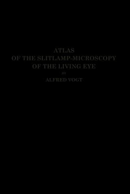
The introduction of the slitlamp of Gullstrand has opened to ophthalmology an entirely new field for clinical observation and diagnosis. It has created what may be . termed an "Histology of the living eye". . Normal and pathologie conditions heretofore established only anatomically can be directly observed in the living eye. lt shows us not only structures that are known, but in addition aseries of observations on histological details, heretofore impossible. These structures, owing partly to their delicacy. , were formerly sacrificed in the process of fixation, or it was impossible to further differentiate them by any method of staining. For instance, we have up to the present failed of anatomical proof of the numerous physiologio remnants of the tunica vasculosa lentis, the arteria hyaloidea, the various intricacies of the framework supporting the vitreous body, types of Jens sclerosis, eto. , but the number of facts already known as a result of anatomical research which have hitherto evaded clinical confirmation, is far greater, The slitlamp, in combination with the corneal microscope, perinits us to observe the living endothelium on the posterior surface of the cornea. Every individual endo- thelial cell on Descemet's membrane, as well as each pathologically deposited lympho- eyte is revealed. The nerve fibres of the cornea can be traced to their very finest ramifications. In Deseemet's and Bowman's membranes we have observed pathological folds,' manifested by their charaeteristic reflexes.
The introduction of the slitlamp of Gullstrand has opened to ophthalmology an entirely new field for clinical observation and diagnosis. It has created what may be . termed an "Histology of the living eye". . Normal and pathologie conditions heretofore established only anatomically can be directly observed in the living eye. lt shows us not only structures that are known, but in addition aseries of observations on histological details, heretofore impossible. These structures, owing partly to their delicacy. , were formerly sacrificed in the process of fixation, or it was impossible to further differentiate them by any method of staining. For instance, we have up to the present failed of anatomical proof of the numerous physiologio remnants of the tunica vasculosa lentis, the arteria hyaloidea, the various intricacies of the framework supporting the vitreous body, types of Jens sclerosis, eto. , but the number of facts already known as a result of anatomical research which have hitherto evaded clinical confirmation, is far greater, The slitlamp, in combination with the corneal microscope, perinits us to observe the living endothelium on the posterior surface of the cornea. Every individual endo thelial cell on Descemet''s membrane, as well as each pathologically deposited lympho eyte is revealed. The nerve fibres of the cornea can be traced to their very finest ramifications. In Deseemet''s and Bowman''s membranes we have observed pathological folds,'' manifested by their charaeteristic reflexes.
Get Atlas of the Slitlamp-Microscopy of the Living Eye by at the best price and quality guranteed only at Werezi Africa largest book ecommerce store. The book was published by Springer-Verlag Berlin and Heidelberg GmbH & Co. KG and it has pages. Enjoy Shopping Best Offers & Deals on books Online from Werezi - Receive at your doorstep - Fast Delivery - Secure mode of Payment
 Jacket, Women
 Woolend Jacket
 Western denim
 Mini Dresss
 Jacket, Women
 Woolend Jacket
 Western denim
 Mini Dresss
 Jacket, Women
 Woolend Jacket
 Western denim
 Mini Dresss
 Jacket, Women
 Woolend Jacket
 Western denim
 Mini Dresss
 Jacket, Women
 Woolend Jacket
 Western denim
 Mini Dresss


